Call us today on: 01233 502314 or 07557 435907

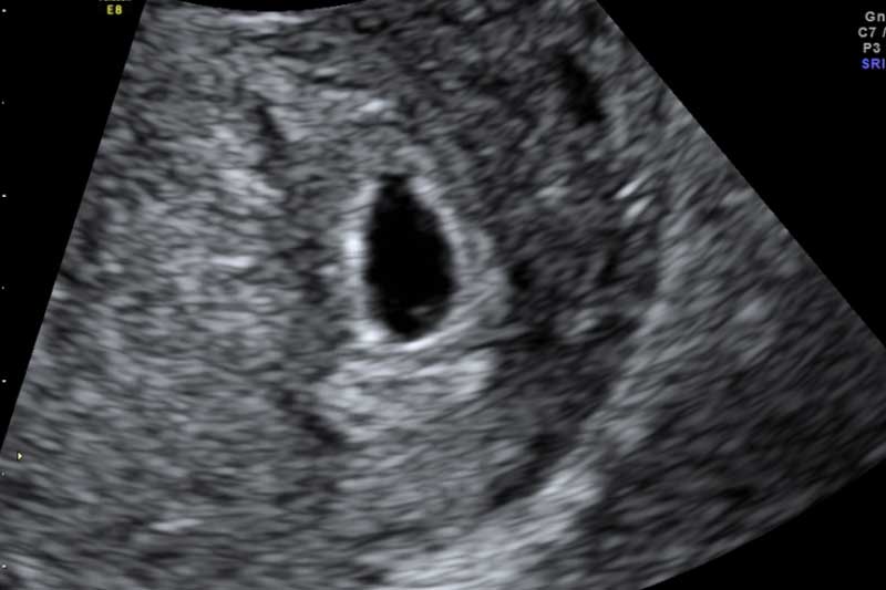
In the provided images, Baby Scan Ashford will illustrate a pregnancy at 5 weeks or more, though clarity may vary among individuals. At this stage, without being able to precisely measure the embryo, we typically rely on assessing the mean sac diameter, which involves three measurements of the sac to estimate a due date. However, this method isn't as precise as measuring the embryo itself.
What can typically be seen is a dark area representing the gestation sac, with a small white circle inside known as the yolk sac. The yolk sac functions to provide nutrients to the developing embryo until the placenta assumes this role later in pregnancy. The presence of only a yolk sac allows us to confirm whether the pregnancy is correctly situated or potentially ectopic.
Moving to a 6-week pregnancy, it becomes feasible to visualise the embryo more clearly and accurately measure the gestational age. However, detecting a heartbeat at this stage isn't always guaranteed.
The absence of a visible heartbeat during this period can lead to anxiety, prompting the need for a follow-up scan in a week or so. To mitigate this uncertainty, at Baby Scan Ashford, we typically recommend early pregnancy scans starting from 7 weeks.
At 7 weeks, the embryo measures approximately 10mm from head to bottom or crown to rump. While the yolk sac might not be visible in all cases, it's usually possible to detect a heartbeat at this stage.
This is why we advocate for early pregnancy scans around this time, as it can alleviate the stress of potential uncertainties and reduce the need for additional appointments.
To summarise the precise differences between 5,6 and 7 weeks of pregnancy scans:
5 Weeks Pregnant:
6 Weeks Pregnant:
7 Weeks Pregnant:
However, it's essential to acknowledge the variability in menstrual cycle lengths, which are typically estimated at 28 days but can range from 21 to 35 days. Consequently, if your cycle is longer or shorter, you may be slightly less or further along in your pregnancy. By aiming for a 7-week scan, we increase the likelihood of capturing all necessary information in one appointment, despite the anticipation felt by many due to the early detection capabilities of today's pregnancy tests.
It's crucial to note that if you experience bleeding or pain, it's essential to rule out the possibility of an ectopic pregnancy. In such cases, a visit to the early pregnancy assessment unit may be necessary, where they can conduct further scans and quantitative Beta hCG measurements to gather additional information. Nonetheless, sometimes a watchful waiting approach might be deemed appropriate. Further details on ectopic pregnancy will be provided later. If you have any questions about any of our scan packages then just get in touch.